
Comparing Error Types and Rates of X-Ray and O-Arm Guided Pedicle Screw Placement
Rishi Vakulabharanam
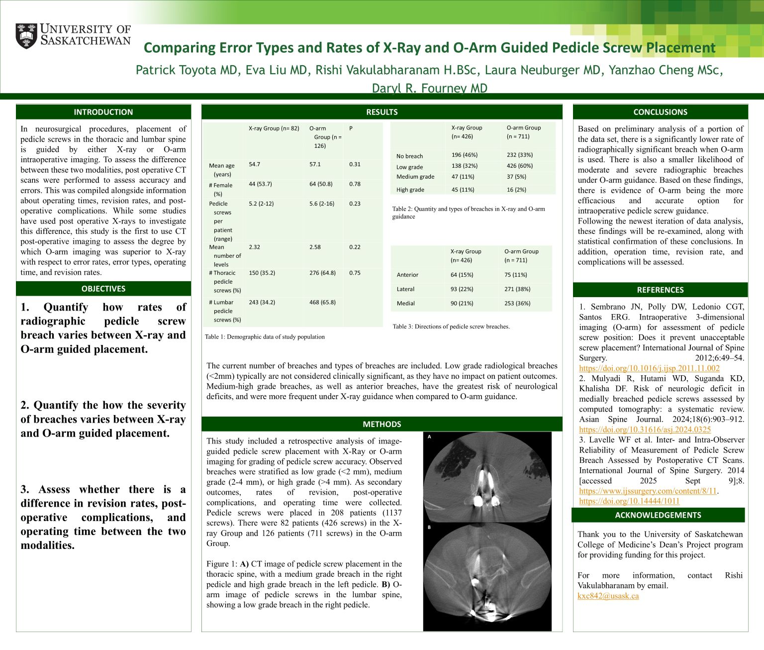
In neurosurgical procedures, placement of pedicle screws is guided by either X-ray or O-arm intraoperative imaging. Different breaches occur during this with respect to distance outside of the pedicle and direction of breach. Medium/high grade breaches (>2 mm) and anterior breaches are the most impactful on patient outcomes. To assess the difference between these two modalities, post-operative CT scans were performed to assess accuracy and errors. This was compiled alongside information about operating times, revision rates, and post-operative complications. This study included a retrospective analysis of 208 patients (1137 screws). There were 82 patients (426 screws) in the X-ray Group and 126 patients (711 screws) in the O-arm Group. Breaches were stratified by direction and severity. X-ray guidance showed a higher rate of anterior breach and medium/high grade breaches, but O-arm guidance showed a higher rate of overall breach, and a higher rate of low-grade breach. However, these low-grade breaches are not associated with an increased risk of neurological deficits. Based on these findings, O-arm guided pedicle screw placement is superior to X-ray guidance with respect to error rates and types. In the future, a larger population will be analyzed for operative time, complication rate, and revision rate.