
3D Shape Modelling Reveals Structural Knee Differences in Aging and Osteoarthritis
Samantha Leech
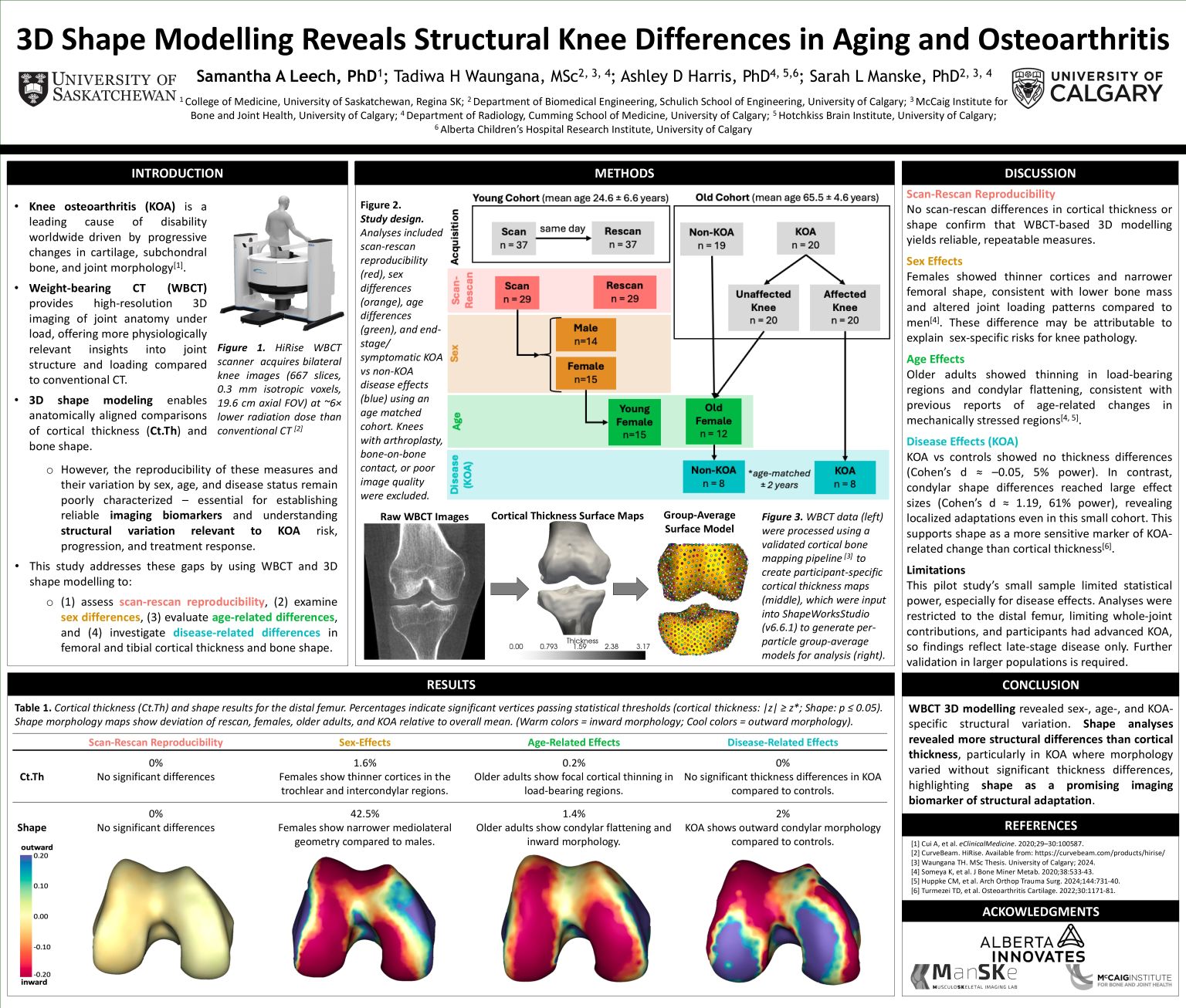
Background: Structural bone adaptation contributes to pain, disability, and progression in knee osteoarthritis (KOA), yet radiographs cannot capture subtle 3D alterations. This study used weight-bearing CT (WBCT), which enables physiologic imaging under load, to quantify cortical thickness and 3D bone shape, complementary metrics of structural adaptation, as potential imaging biomarkers for KOA risk, progression, and treatment response.
Methods: Cortical thickness maps, generated with a validated cortical bone mapping pipeline, were aligned to WBCT surface masks and loaded into ShapeWorks for statistical shape modelling and cortical thickness network analyses. Analyses assessed scan-rescan reproducibility in young adults (n=29), sex differences (15 females, 14 males), age effects (15 young adults vs 19 older females), and disease effects (KOA n=8 vs age-matched controls n=8). Knees with arthroplasty, bone-on-bone contact, or poor image quality were excluded.
Results: WBCT-based modelling was reproducible. Females had thinner cortices in the trochlear and intercondylar regions and narrower mediolateral geometry. Older adults showed cortical thinning in load-bearing regions with condylar flattening. KOA cases exhibited no cortical thinning but outward condylar morphology.
Conclusion: Shape analyses revealed more structural differences than cortical thickness, particularly in KOA where morphology varied without significant thickness changes, highlighting shape as a promising imaging biomarker of structural adaptation.