
Time-Lapsed 4D Synchrotron Imaging of early PTH-induced Changes in Trabecular Bone of the Rabbit Calcaneus
Ali Rizvi
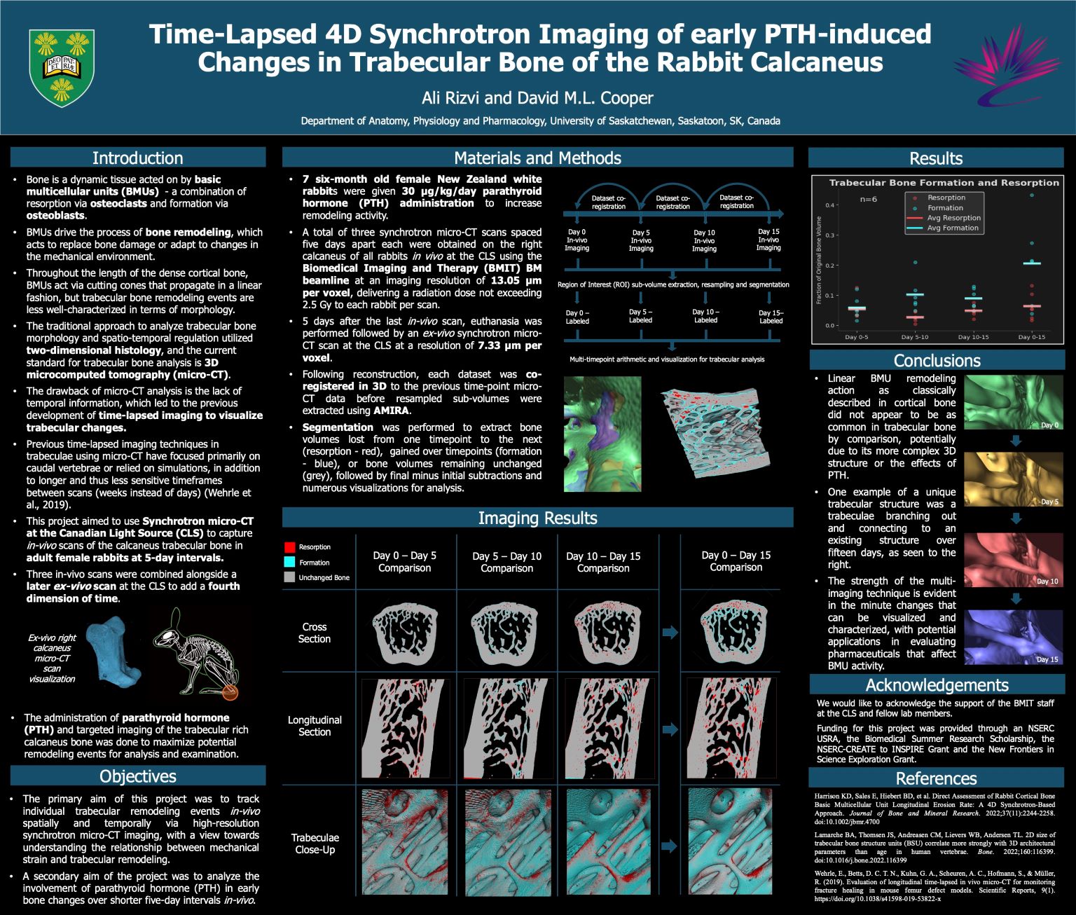
The overarching goal of this project was to understand the relationship between mechanical strain and trabecular bone remodeling via time-lapsed imaging techniques, as the complex three-dimensional nature of trabecular bone remodeling has traditionally been less well-defined than linear remodeling in dense cortical bone. Using synchrotron microcomputed tomography (micro-CT), this project sequentially imaged calcanei from female New Zealand white rabbits to track individual trabecular bone changes with three in vivo scans and one ex vivo scan spaced five days apart each. The rabbits were administered 30 µg/kg/day of parathyroid hormone (PTH) over these fifteen days to increase remodeling activity for later examination, with another project aim being to understand the role of PTH in early bone morphology changes in vivo. After imaging, the successful registration of each scan and subsequent visualizations established the success of the multiple time point imaging technique, with observed examples including trabecular rods increasing in length and gradual resorption over trabecular surfaces. Our results also showed a significant increase in bone volume fraction in the trabecular region over fifteen days of PTH administration (p=0.026). With these results, our next aim moving forward will be to focus on better understanding the relationship between mechanical strain and trabecular bone remodeling.