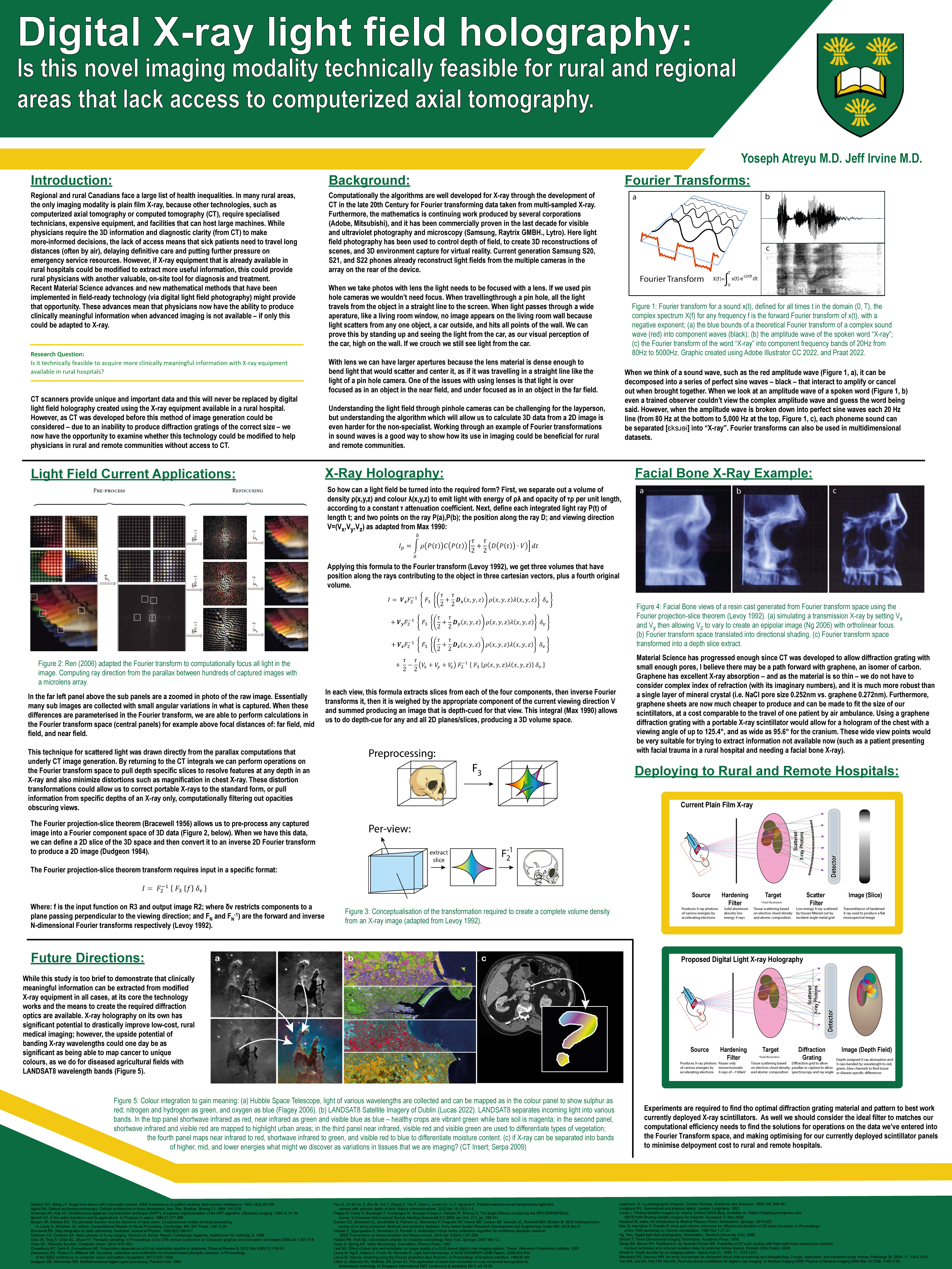
B1.5: Digital X-ray light field holography: Is this novel imaging modality technically feasible for rural and regional areas that lack access to computerized axial tomography
Yoseph Atreyu, Jeff Irvine
Regional and rural Canadians face a large list of health inequalities, this project is trying to address whether currently deployed X-ray equipment, with minimal adaptation, could provide more clinical meaning when advanced imaging is not feasible. Rural populations and especially remote populations do not have equal access to advanced imaging modalities. CT is an expensive imaging modality requiring special equipment, and radio shielded MRI even more so. Remote patients needing access to these methods must travel long distances. However, X-ray is cheaper and can potentially go anywhere. Can we extract more information from currently deployed plain film X-ray equipment to address equitable access for rural and remote patients? This is a desktop study where we review currently deployed X-ray and light field technology to evaluate whether light field imaging could apply to X-ray image acquisition in rural and remote Canada. (Bio ID: 3073) This study does not constitute research involving humans and is thus exempt from Research Ethics Board (REB) review and approval. Light field acquisition appears to be feasible if wavelength matched diffraction grating can be created. Further, the light field algorithm can be adapted to solve for wavelength which could give additional information currently filtered out of X-ray and CT image capture.
Initially this study investigated whether our X-ray equipment would be able to calculate 3D volume information; additionally, as the light field calculation can extract “colour” from X-ray wavelengths and this may potentially reveal additional information not available otherwise. For diagnostic imaging 360° computer tomography will always remain a mainstay, however this study shows that with suitable X-ray diffraction grating a single plain film X-ray may allow 3D data and wavelength data which could potentially represent a new mode of medical imaging. We recommend testing existing software presets (MATLAB) against image capture with various diffraction gratings with and without linear scatter filters to find the optimal diffraction grating for medical imaging, and further we recommend collaboration with the optics community to further optimize the calculations of light fields for currently available diffraction gratings with X-ray.
