
Establishing a Protocol for Visualizing Lipid Droplets in Rat Gut Enterocytes
Ario Safaeian
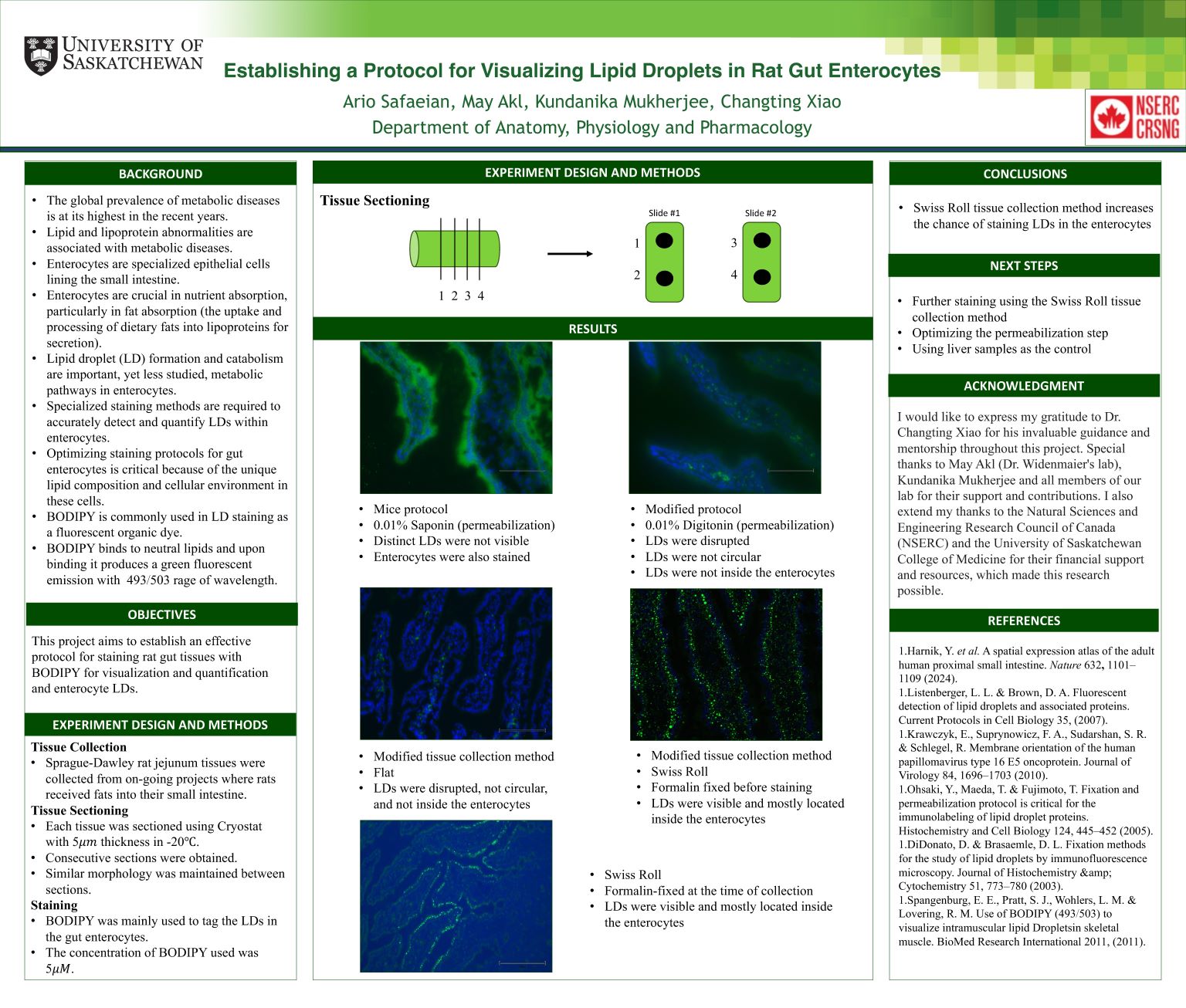
Dysregulated lipid processing in intestinal absorptive cells (Enterocytes) leads to lipid and lipoprotein abnormalities accompanied by an increased risk of cardiovascular disease in patients. This project aims to establish a protocol for visualizing enterocyte LDs with BODIPY staining in rat intestinal tissues. Jejunum tissue was collected after the rats were provided with lipids into the small intestine lumen and sectioned using Cryostat at -20 with 5 thickness. Using a protocol adopted from mice, LDs seemed disrupted, dislocated or not clearly visible. Further, we tested several modifications to this protocol, including with and without permeabilization (with various reagents, e.g. Saponin, Tween 20, and Digitonin), sample collection and fixation methods. Preliminary results indicate that permeabilization did not disrupt the LDs. Clear LD images were obtained with tissues collected using the Swiss Roll (SR) method with fixation at the time of collection. The stained LDs in SR samples were mostly contained and not disrupted. Furthermore, their locations in the intestinal tissue and enterocytes were consistent with LD biology. Further refining of the protocol is ongoing. An optimal protocol for visualizing rat enterocyte LDs with BODIPY staining would likely include the collection of intestinal tissues with the SR method along with fixation at the time of collection and with optional permeabilization.