
Bedside ultrasound changing emergency medicine
Point of Care Ultrasound helping to change bedside diagnosis
By Marg SheridanWhile the public has seen the size of cell phones shrink from their Zack Morris ‘brick’ days in the 1990’s, medical professionals have witnessed a similar ‘miniaturization’ of the equipment used in ultrasound imaging.
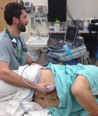
For Dr. Paul Olszynski, the smaller Point of Care Ultrasound (POCUS) machines represent one of those improvements that can make diagnosis easier for emergency doctors, and less stressful and time-consuming for patients.
“POCUS is defined as an ultrasound being done by the clinician right at the bedside,” Olszynski explained. “It is usually very focused – often answering a binary question or two rather than being comprehensive like the scans done by our colleagues in Medical Imaging.
“So it means knowing tremendously more about the patient at the bedside.”
It’s a change that Olszynski said has taken time in that the technology needed to first become more available in terms of size and ease of use, while at the same time required doctors to start thinking about a new way of applying the tool.
“Right now you can have machines much more powerful than even the biggest machines from 20-30 years ago,” Olszynski continued. “You now have those in the size of a laptop - so when you have that kind of power, with that kind of image resolution, it really helps advance the field.”
POCUS has enabled doctors to recognize injuries and illnesses ranging from bleeding in the abdomen from trauma, to gallstones, kidney obstruction, and even acute heart failure right at the bedside. And using POCUS to identify these problems in the emergency room has led to an ever-expanding list of uses for the tool.
“(We) started to say ‘well, let’s look at things that haven’t been looked at with ultrasound,’” Olszynski continued. “So let’s look at the lungs and see if we can determine whether or not someone has a pneumothorax, and it turns out it’s a phenomenal test – far better than an x-ray for identifying pneumothorax.”
He’s now seeing POCUS being used to assist with the diagnose of a range of issues from ocular disorders to bowel obstructions, but that doesn’t mean that the team he works with have stopped trying to expand on the ways POCUS can help to make a difference. They’re currently looking at the role ultrasound can play in diagnosis of pediatric pneumonia, and how lung ultrasounds in heart failure patients can help to determine not only their response to therapy, but better predict discharge.
They’ve also recently completed a chart review of 400 POCUS assessments in the emergency room with an emphasis on image concordance and impact on care – so whether what the emergency doctor was seeing with POCUS was consistent with the other imaging the patients received further along.
In addition to the clinical research, one of Olszynski’s focuses is also how to incorporate POCUS into the medical curriculum at the College of Medicine.
“Introducing POCUS to the undergrad curriculum serves two purposes: the primary one is to serve the curriculum,” Olszynski said. “(Meaning) there’s lots of evidence suggesting that ultrasound can facilitate or improve the way you teach the traditional content.
“The secondary goal is, given that POCUS is becoming a very common skill throughout pretty much every specialty – internal medicine, general surgery, emergency medicine, intensive care, anesthesia – it makes sense to introduce the students to it at an undergrad level.”
They’ve already introduced ultrasound into the first-year anatomy course, so students get the opportunity to learn how to generate and interpret ultrasound images along with what they’re learning in their cadaveric lab and in other course work. Olszynski stressed that this use of ultrasounds helps to complement and clarify some of the anatomy and applied anatomy concepts.
“Ultrasound is dynamic, so they can actually see what it means for the lung - during inspiration - to move down, and then come back up during expiration,” he continued. “They can see what the heart looks like when it’s beating in real time - so they can see the valves opening and closing coinciding with the ventricles contracting and filling.
“So it’s serving its purpose to facilitate the curriculum, and it’s a more dynamic, and more hands-on way of learning.”
And as Olszynski works to help educate students in some of the newer uses for POCUS, he’ll be opening the door to the possibility that those student will one day also find a new use for the tool which can help to change the way patients are diagnosed and treated.
“It’s a transition, and it’s an important one – and the reality is that that needs to start sooner than residency.”