
Imaging Platform helping create ‘Google-map’ of cancer, grow cartilage
Dr. Franco Vizeacoumar and his team are working on building a ‘Google-map’ of cancer cells with the help of a high-tech robotic imaging platform
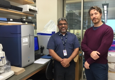
By Marg SheridanIn a fourth-floor lab in the Health Sciences Building Dr. Franco Vizeacoumar and his team are working on building a ‘Google-map’ of cancer cells with the help of a high-tech robotic imaging platform – a platform that won him, and research partner Dr. Brian Eames, $500,000 in funding from the Canadian Foundation for Innovation (CFI).
“We want to know what the pathways are that drive tumour formation – and to do this we need to build a roadmap,” explained Vizeacoumar. “Just like Google maps tell us which streets connect location A to B, and how we can intersect these pathways to selectively block them.”
The hope is that the map would then help in figuring out where the cellular mutation occurs that leads to the ‘turning-on’ of the cancer.
“If we can develop the hi-resolution maps, just like the street maps, then we’ll know who the ultimate culprit is,” Vizeacoumar continued. “(Then we) know where we have to target, and how we can kill the cancer cells alone.”
It’s an arduous task, and if done conventionally the time it would take the team to look at the cells individually under a microscope would be enormous. With an estimated ~20,000 genes in the human genome, Vizeacoumar estimates he’d be working on the task for about 120,000 weeks just to collect the data for a single genetic or environmental perturbation.
This is where his High Throughput Molecular Imaging Platform steps in.
“It’s image-based,” Vizeacoumar said. “You can actually look at the still and see how the (cells) are changing - and are we really killing them.
“So we’ve generated this quantitative data that we can actually go after and develop therapeutic targets.”
And while Vizeacoumar has been looking at how chromosome changes can influence cancer cell death, Eames, an Assistant Professor in Anatomy and Cell Biology, has been using the platform to continue his research into cartilage formation.
“There are ~20,000 genes in most vertebrates, so potentially that would take a lot of time to figure out the function of each particular gene and how it’s relevant to disease,” explained Eames. “This equipment has the ability to test each of these individual genes in a screen, you don’t even have to know what each gene is doing at the molecular level.
“The only thing you know is activating that gene will make it, in my case, turn into bone or cartilage better.”
Eames’ studies will also use the imaging platform to assess gene function in living zebrafish embryos, an application he believes is the first of its’ type, and can have a wide-range of applications including the ability to create bone and cartilage using the host’s cells.
“Bone fractures, osteoporosis, osteoarthritis - those are all clinical conditions that can really benefit from understanding how you get cells to turn into bone and cartilage,” Eames stressed. “This high-throughput approach has been around for a while, you use robots to manipulate cells, and to perform an assay to show what changes happen to the cells happens.
“But the really cool part that blew my mind is that our equipment has an added imaging analysis component (that) takes a picture of each well to document the changes to the cell after the high-throughput genetic and environmental manipulations, and then you program the software to analyze the results.”
The platform allows them to tell the analytical software what they’re looking for - the specific parameters they’re interested in measuring - which allows the computer to analyze the data, do the assays, and produce the refined data for any project. However, even after it has parsed through the results, the data created is immense and requires a computational team and analysis experts to sift through.
Vizeacoumar and Eames seem like unlikely partners based solely on their research subjects alone, however while their practical research goals differ, the imaging platform’s versatility created a bridge for interdisciplinary research.
“Franco is interested in cancer, I’m interested in bone and cartilage – those differences turned out not to be that important,” Eames continued. “What we shared was this knowledge of cell biology, these basic cell biology questions, and an appreciation of how this equipment can move forward with answering the questions. This imaging platform throws me, and the basic biomedical sciences at U of S, into a whole different level of cutting-edge research.”
The CFI funding Vizeacoumar and Eames have received came courtesy of the John R. Evans Leaders Fund, designed to help universities attract, and retain, leading researchers.