
Combining medicine and engineering to repair a damaged heart
Regenerating heart muscle tissue using a 3D printer—once the stuff of Star Trek science fiction—now appears to be firmly in the realm of the possible
By University CommunicationsThe combination of the Canadian Light Source (CLS) synchrotron’s unique biomedical imaging and therapy (BMIT) beamline and the vision of a multi-discipline researcher from the University of Saskatchewan in confirming fiction as fact was published in the September issue of Tissue Engineering, one of the leading journals in this emerging global research field of tissue regeneration.
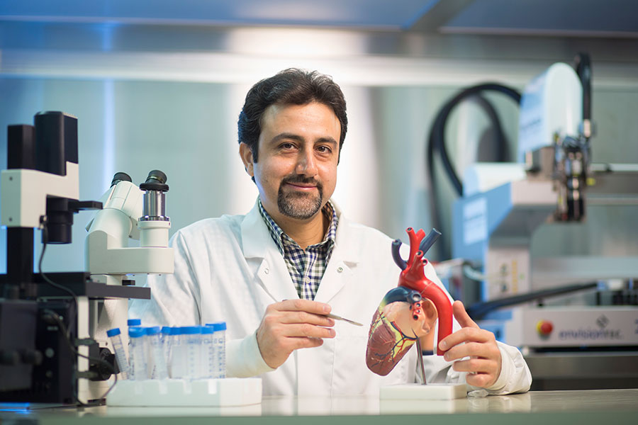
U of S researcher Mohammad Izadifar says he is combining medicine and engineering to develop ways to repair a damaged heart.
“The problem is the heart cannot repair itself once it is damaged due to a heart attack.” he explained.
Izadifar has conducted his research out of three places on campus: the College of Engineering, the CLS and the College of Medicine where he has been certified in doing open heart surgery on rats, having trained in all the ethical protocols related to these research animals.
And thanks to the confirmation photo images he has from his collaboration with the CLS, Izadifar has already proven the 3D printed human cells, which he has dubbed the “heart patch,” can start to grow as intended in theory.
Once implanted in the laboratory mice, the heart patch is invisible to regular medical imaging. Izadifar has developed an X-ray imaging technique at the CLS to monitor the 3D-printed heart patch after implanting them in the laboratory mice. The CLS-derived pictures submitted to the journal show a 3D-printed heart patch with human cells arranged in 200 micron-wide strands with the distance between each strand being 400 microns. One micron is one-thousandth of a millimeter.
Izadifar says the key in printing live human tissue is finding the right gel medium to become the “ink” for the printer.